Knee Arthroscopy
Introduction
Physiotherapy in Edmonton for Knee
Welcome to Family Physiotherapy’s Guide to Knee Arthroscopy.
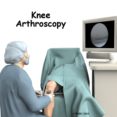 The use of arthroscopy has revolutionized many different types of orthopedic surgery. During arthroscopy, a small video camera attached to a fiber-optic lens is inserted into the body to allow a physician or surgeon to see inside without making a large incision (arthro means joint, scopy means look). The knee was the first joint in which the arthroscope was commonly used to both diagnose problems and to perform surgical procedures inside a joint.
This guide will help you understand:
- what parts of the knee are involved
- what types of conditions can be treated
- what to expect after surgery
- what Family Physiotherapy’s approach to rehabilitation is
Anatomy
What parts of the knee are involved?
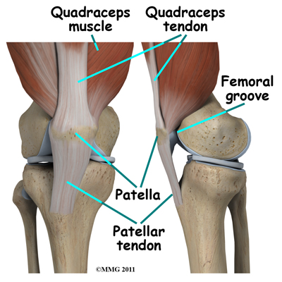
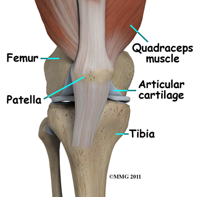 The knee joint is formed where the femur (lower end of the thighbone) connects with the tibia (upper end of the main lower leg bone). On the front of the joint is the patella (kneecap). The patella is what is called a sesamoid bone that is a part of the extensor mechanism of the knee joint. The extensor mechanism connects the large muscles of the thigh to the tibia such that contracting the thigh muscles pulls on the tibia and allows us to straighten the knee. The parts of the extensor mechanism include the thigh muscles, the quadriceps tendon, the patella and the patellar tendon.
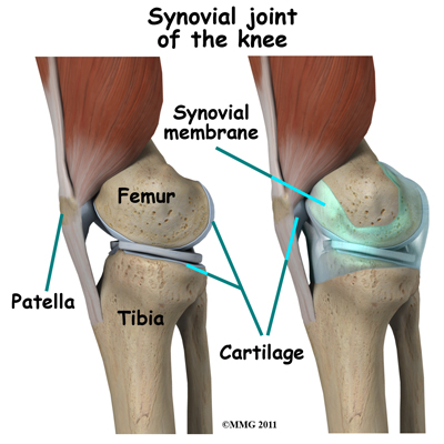
The knee joint is surrounded by a water-tight pocket called the joint capsule.
This capsule is formed by the knee ligaments, connective tissue, and synovial tissue. When the joint capsule is filled with sterile saline and is distended, the surgeon can insert the arthroscope into the pocket that is formed, turn on the lights and the camera, and see inside the knee joint as if looking into an aquarium. The surgeon can see nearly everything that is inside the knee joint including: (1) the joint surfaces of the tibia, femur and patella, (2) the two menisci, (3) the two cruciate ligaments, and (4) the synovial lining of the joint.
 There is one meniscus on each side of the knee joint. The C-shaped medial meniscus is on the inside part of the knee, closest to your other knee (medial means closer to the middle of the body.) The U-shaped lateral meniscus is on the outer half of the knee joint (lateral means further out from the center of the body.)
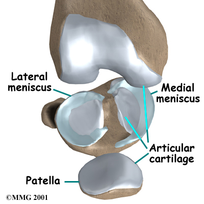 The menisci (plural for meniscus) protect the articular cartilage on the surfaces of the thighbone (femur) and the shinbone (tibia) and help to create a deeper joint surface, which aids in joint stability. Articular cartilage is the smooth, slippery material that covers the ends of the bones that make up the knee joint, as well as most other joints. The articular cartilage allows the joint surfaces to slide against one another without damage to either surface.
Ligaments are tough bands of tissue that connect the ends of bones together and help to keep the bones together, creating a stable joint. The anterior cruciate ligament (ACL) is located in the center of the knee joint where it runs from the backside of the femur, or thighbone’s joint surface to connect to the front of the tibia, or shinbone’s joint surface.
The ACL runs through a special notch in the femur called the intercondylar notch and attaches to an area of the tibia called the tibial spine.
The ACL is the main controller of how far forward the tibia moves under the femur. This is called anterior translation of the tibia. If the tibia moves too far, the ACL can rupture. The ACL is also the first ligament that becomes tight when the knee is straightened. If the knee is forced past this point, or hyperextended, the ACL can also be torn.
The posterior cruciate ligament (PCL) is located near the back of the knee joint. It also attaches to the femur’s joint surface and the tibia’s joint surface, but crosses behind the ACL such that the two ligaments (the ACL and PCL) form somewhat of an X inside the joint (the term ‘cruciate’ means shaped like a cross.)
The PCL is the primary stabilizer of the knee and the main controller of how far backward the tibia moves under the femur. This motion is called posterior translation of the tibia. If the tibia moves too far back, the PCL can rupture.
Related Document: Family Physiotherapy's Guide to Knee Anatomy
Rationale
What does my surgeon hope to accomplish?
When knee arthroscopy first became widely available in the 1970's it was used primarily to look inside the knee joint and make a diagnosis. Today, knee arthroscopy is used in performing a wide range of different types of surgical procedures on the knee joint including confirming a diagnosis, removing loose bodies, removing or repairing a torn meniscus, reconstructing torn ligaments, repairing articular cartilage and fixing fractures of the joint surface.
Your surgeon's goal is to fix or improve your problem by performing a suitable surgical procedure; the arthroscope is a tool that improves the surgeon’s ability to perform that procedure. The arthroscope image is magnified and allows the surgeon to see better and clearer, and perform surgery using much smaller incisions than with traditional surgery. This results in less tissue damage to normal tissue and can shorten the healing process. Remember, however, that the arthroscope is only a tool. The results that you can expect from a knee arthroscopy depend on what is wrong with your knee, what can be done inside your knee to improve the problem, as well as your effort at rehabilitation after the surgery.
Preparations
What do I need to know before surgery?
You and your surgeon should make the decision to proceed with surgery together. Sometimes arthroscopic surgery is done as an immediate treatment for an injury, such as in the case of a knee with a torn meniscus that limits the knee from fully straightening. Sometimes, however, arthroscopic surgery is performed as a last resort to treatment, for example if physiotherapy treatment combined with a home exercise program has failed to ease the pain of a worn down patellar joint surface, an arthroscopic surgery may be suggested.
You need to understand as much as possible about the surgical procedure. If you have concerns or questions, be sure and talk to your surgeon.
Once you decide on surgery, you need to take several steps. Your surgeon may suggest a complete physical examination by your regular doctor. This exam helps ensure that you are in the best possible condition to undergo the operation.
If you have not already tried physiotherapy for your injury, you should spend time at Family Physiotherapy before the surgery with the physiotherapist who will be managing your rehabilitation after surgery. This allows you to get a head start on your recovery. One purpose of this preoperative visit is to record a baseline of information. Your therapist will check your current pain levels, your ability to do specific activities, and the movement and strength of each knee.
A second purpose of the preoperative visit to Family Physiotherapy is to prepare you for surgery. Your therapist will teach you how to walk safely using crutches or a walker and you will begin learning some of the exercises you'll use during your recovery.
On the day of your surgery, you will probably be admitted for surgery early in the morning. You shouldn't eat or drink anything after midnight the night before.
Family Physiotherapy provides services for physiotherapy in Edmonton.
Preparations
What happens during the procedure?
Before surgery you will be placed under either general anesthesia or a type of spinal anesthesia. In simple cases, local anesthesia may be adequate. Special braces are attached to the operating room table, which are used to safely cradle the leg and allow the surgeon to move the leg and bend the knee easily. Finally, sterile drapes are placed to create a sterile environment for the surgeon to work. There is a great deal of equipment that surrounds the operating table including the TV screens, cameras, light sources and surgical instruments.
The surgeon begins the operation by making two or three small openings into the knee, called portals. These portals are where the arthroscope and surgical instruments are placed inside the knee. Care is taken to protect the nearby nerves and blood vessels. A small metal or plastic tube (or cannula) will be placed through one of the portals to inflate the knee with sterile saline, which allows the tissues inside the joint to be more easily visible.
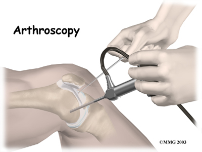
The arthroscope is a small fiber-optic tube that is used to see and operate inside the joint. It is a small metal tube about 1/4 inch in diameter (slightly smaller than a pencil) and about seven inches in length. The fiber-optics inside the metal tube of the arthroscope allow a bright light and a TV camera to be connected to the outer end of the arthroscope. The light shines through the fiber-optic tube and into the knee joint. A TV camera is attached to the lens on the outer end of the arthroscope. The TV camera projects the image from inside the knee joint onto a TV screen next to the surgeon. The surgeon actually watches the TV screen (not the knee joint) while moving the arthroscope to different places inside the knee joint.
Over the years since the invention of the arthroscope, many very specialized instruments have been developed to perform different types of surgery using the arthroscope. Due to these inventions many surgical procedures that once required large incisions for the surgeon to see and fix the problem can now be done with much smaller incisions. For example, simple removal of a torn meniscus or loose body can be done using two small incisions that are approximately 1/4 inch or 6mm. More extensive surgical procedures such as ligament reconstruction or fracture repair may require larger incisions, however, they are still much smaller incisions than what was needed prior to the invention of the arthroscope.
Once the surgical procedure is complete, the arthroscopic portals and surgical incisions will be closed with sutures or surgical staples. Small pieces of surgical tape are also applied to assist the skin to heal as smoothly as possible. Often a large bandage is applied from mid thigh to the toes. Wrapping the entire leg with a compressive bandage reduces swelling and helps prevent blood clots in the leg. Once the bandage has been placed, you will be taken to the recovery room.
Surgical Procedure
What happens during the procedure?
Before surgery you will be placed under either general anesthesia or a type of spinal anesthesia. In simple cases, local anesthesia may be adequate. Special braces are attached to the operating room table, which are used to safely cradle the leg and allow the surgeon to move the leg and bend the knee easily. Finally, sterile drapes are placed to create a sterile environment for the surgeon to work. There is a great deal of equipment that surrounds the operating table including the TV screens, cameras, light sources and surgical instruments.
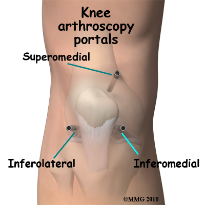 The surgeon begins the operation by making two or three small openings into the knee, called portals. These portals are where the arthroscope and surgical instruments are placed inside the knee. Care is taken to protect the nearby nerves and blood vessels. A small metal or plastic tube (or cannula) will be placed through one of the portals to inflate the knee with sterile saline, which allows the tissues inside the joint to be more easily visible.
The arthroscope is a small fiber-optic tube that is used to see and operate inside the
joint. It is a small metal tube about 1/4 inch in diameter (slightly smaller than a pencil) and about seven inches in length. The fiber-optics inside the metal tube of the arthroscope allow a bright light and a TV camera to be connected to the outer end of the arthroscope. The light shines through the fiber-optic tube and into the knee joint. A TV camera is attached to the lens on the outer end of the arthroscope. The TV camera projects the image from inside the knee joint onto a TV screen next to the surgeon. The surgeon actually watches the TV screen (not the knee joint) while moving the arthroscope to different places inside the knee joint.
Over the years since the invention of the arthroscope, many very specialized instruments have been developed to perform different types of surgery using the arthroscope. Due to these inventions many surgical procedures that once required large incisions for the surgeon to see and fix the problem can now be done with much smaller incisions. For example, simple removal of a torn meniscus or loose body can be done using two small incisions that are approximately 1/4 inch or 6mm. More extensive surgical procedures such as ligament reconstruction or fracture repair may require larger incisions, however, they are still much smaller incisions than what was needed prior to the invention of the arthroscope.
 Once the surgical procedure is complete, the arthroscopic portals and surgical incisions will be closed with sutures or surgical staples. Small pieces of surgical tape are also applied to assist the skin to heal as smoothly as possible. Often a large bandage is applied from mid thigh to the toes. Wrapping the entire leg with a compressive bandage reduces swelling and helps prevent blood clots in the leg. Once the bandage has been placed, you will be taken to the recovery room.
After Surgery
What happens after surgery?
Knee arthroscopy is usually done on an outpatient basis meaning that patients go home the same day as the surgery. More complex ligament reconstructions that require larger incisions and surgery that alters bone may require a short stay in the hospital to control pain more aggressively, monitor the situation more carefully, and to begin physiotherapy.
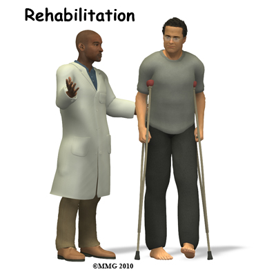 As mentioned above, the portals are covered with surgical strips. The larger incisions may have been repaired with either surgical staples or sutures and the knee may be wrapped in an elastic bandage. Crutches are commonly used after knee arthroscopy even though they may only be needed for one to two days after a simple procedure.
Patients who have had more complex reconstructive surgery may need to wear a knee brace for several weeks. The brace helps to protect the healing tissue inside the knee joint. You may be allowed to remove the brace at times during the day to do gentle range-of-motion exercises and to have a bath.
It is crucial that you follow your surgeon's instructions about how much weight to place on your foot while standing or walking. Most often you are allowed to weight bear as tolerated, but there are instances where your surgeon may request that you do not fully weight bear, such as in the case of a joint surface bone repair. You may be instructed to use a cold pack on the knee and to keep your leg elevated and supported.
Complications
What might go wrong?
As with all major surgical procedures, complications can occur during knee arthroscopy. This document doesn't provide a complete list of the possible complications, but it does highlight some of the most common problems, which are:
- anesthesia complications
- thrombophlebitis
- infection
- equipment failure
- slow recovery
Anesthesia Complications
Most surgical procedures require that some type of anesthesia be done before surgery. A very small number of patients have problems with anesthesia. These problems can be reactions to the specific drugs used, problems related to other medical complications, and problems due to being under anesthesia. Be sure to discuss the risks of anesthesia and any specific concerns related to your own health or pre-existing conditions with your anesthesiologist.
Thrombophlebitis (Blood Clots)
Thrombophlebitis, sometimes called deep venous thrombosis (DVT), can occur after any operation, but they are more likely to occur following surgery on the hip, pelvis, or knee. DVT occurs when blood clots form in the large veins of the leg. This may cause the leg to swell and become warm to the touch and painful. If the blood clots in the veins break apart, they can travel to the lungs, where they lodge in the capillaries and cut off the blood supply to a portion of the lung. This is called a pulmonary embolism. (Pulmonary means lung, and embolism refers to a fragment of something traveling through the vascular system.) Most surgeons take the risk of DVT and preventing it very seriously. There are many ways to reduce the risk of DVT, but probably the most effective is to get you moving as soon as possible after surgery. Two other commonly used preventative measures include the use of pressure stockings to keep the blood in the legs moving, and medications that thin the blood and prevent blood clots from forming in the first place.
Infection
Following knee arthroscopy, it is possible that a postoperative infection may occur. Fortunately getting an infection is very uncommon and happens in less than 1% of cases. If, however, you do get a postoperative infection you may experience symptoms such as increased pain, swelling, fever and a redness or drainage from the incisions.
Infections are of two types: superficial or deep. A superficial infection may occur in the skin around the incisions or portals. A superficial infection does not extend into the joint and can usually be treated with antibiotics alone. If the knee joint itself becomes infected, this is a serious complication and will require antibiotics and possibly another surgical procedure to drain the infection. You should alert your surgeon immediately if you think you are developing an infection.
Equipment Failure
Many of the instruments used by the surgeon to perform knee arthroscopy are small and fragile. These instruments can be broken resulting in a piece of the instrument floating inside the knee joint. The broken piece is usually easily located and removed, but this may cause the operation to last longer than planned. There is usually no damage to the knee joint due to the breakage.
Different types of surgical devices (screws, pins, and suture anchors) are used to hold tissue in place during and after arthroscopy. These devices can also cause problems. If one breaks, the free-floating piece may hurt other parts inside the knee joint, particularly the articular cartilage. Another issue may be that the end of a tissue anchor pokes too far through the tissue and may rub and irritate other nearby tissues. A second surgery may be needed to remove the device or fix problems associate with failure of these devices.
Slow Recovery
Not everyone gets quickly back to routine activities after knee arthroscopy. Because the arthroscope allows surgeons to use smaller incisions than in the past, many patients mistakenly believe that less surgery was necessary, however, this is not always true. The arthroscope allows surgeons to do a great deal of reconstructive surgery inside the knee without making large incisions. How fast you recover from knee arthroscopy depends on what type of surgery was done inside your knee. Simple problems that require simple procedures using the arthroscope generally get better faster. Patients with extensive damage to the knee ligaments or articular cartilage tend to require more complex and extensive surgical procedures. These more extensive reconstructions take longer to heal and have a slower recovery. You should discuss this with your surgeon and make sure that you have realistic expectations of what to expect following arthroscopic knee surgery. How fast you recover also depends on how compliant you are to your individual rehabilitation program. If you have difficulty completing the exercises that your physiotherapist at Family Physiotherapy has prescribed for you, you should discuss this with them. Modifications to your program can be made to ensure that although recovery may not be as quick as it could be by completing the entire rehabilitation program, you will at least always be moving in the forward direction with regards to improvement of your surgical knee.
Rehabilitation
What will my recovery be like?
Rehabilitation after knee arthroscopy should begin as soon as possible at Family Physiotherapy once you are discharged from the hospital. There are a few cases where your surgeon may delay a start in your therapy because they want the tissues to heal with rest before any further stress is placed on the joint. Each surgeon will set his own specific restrictions regarding when to begin treatment at Family Physiotherapy based on what was done during the surgical procedure, their personal experience, and whether your tissues are healing as expected. In regards to overall recovery time, generally speaking, the more complex the surgery, the more involved and prolonged your rehabilitation program will be.
If you are still using crutches by the time we first see you at Family Physiotherapy, your physiotherapist will ensure you are using the crutches safely, properly, and confidently and that you are abiding by your weight bearing restrictions if you have been given any. We will also ensure that you can safely use your crutches on stairs. If you are no longer using crutches, or once you no longer need them, your physiotherapist will focus on normal gait re-education so you are putting only the necessary forces through the surgical side with each step, and are not compensating in any way. Until you are able to walk without a significant limp, we recommend that you continue to use your crutches, or at least one crutch or a cane/walking stick. Improper gait can lead to a host of other pains in the knee, hip and back so it is prudent to use a walking aid until near normal walking can be achieved. Your Family Physiotherapy physiotherapist will advise you regarding the appropriate time for you to be walking without any walking aid at all.
During your first few appointments at Family Physiotherapy your physiotherapist will focus on relieving any pain and inflammation that you may still have from the surgical procedure itself. We may use modalities such as ice, heat, ultrasound, or electrical current to assist with decreasing any pain or swelling you have around the surgical site or anywhere down the leg. In addition, your physiotherapist may massage your leg and ankle to improve circulation and help decrease your pain.
The next part of our treatment will focus on regaining the range of motion in your knee. Your physiotherapist at Family Physiotherapy will prescribe a series of exercises that you will practice in the clinic and also learn to do as part of a home exercise program. Range of motion in the knee generally comes back very quickly after an arthroscopic surgery, but it still depends on what your surgeon has done inside your joint, how much swelling is present, and how controlled your discomfort is. More complicated reconstructive surgeries will take longer to regain range of motion. During the exercises you may experience a small amount of discomfort at the end ranges of motion initially. Despite this discomfort it is still important to perform the range of motion exercises prescribed because moving the joint also helps to move the swelling, get fresh blood to the healing area, and provide nutrition to the surface of the joint. Only mild discomfort, however, is permissible. Any sharp or moderate discomfort should be heeded. An exercise bike at this stage of your rehabilitation is very useful to assist in gaining back the knee range of motion. Even if you are unable to fully rotate the pedals of the bike using it is still encouraged. Performing the simple back and forth motion forces fluid through the joint, which helps to move the swelling and bring fresh blood to the healing tissues.
One goal after an arthroscopic surgery is to regain full bending and straightening of your knee joint. Without this full range of motion, areas of the joint surface cartilage can become weak and start to wear down. In addition, without full range of motion the biomechanics of the knee do not function as they have been designed to, and this contributes to early wearing down of the joint. Often prior to arthroscopic surgery fully extending or bending the knee is not possible and the goal of the surgery in the first place is to deal with any issues inside the joint that are limiting this motion. For this reason, your physiotherapist at Family Physiotherapy will be strict in encouraging you to regain both the full bending and straightening of the knee joint. There are a few limited instances where the knee joint will not regain its full range of motion even after an arthroscopic surgery and this limited range of motion in these few cases is accepted.
These instances, however, are rare and your surgeon will inform you if your knee is one of these exceptions. In all other cases, it is crucial that you regain the full bending and straightening of your knee in order to maximize the functioning of your knee and avoid further problems in the future.
In addition to you yourself doing range of motion exercises your physiotherapist may mobilize your knee joint to assist in regaining motion. This hands-on technique encourages the knee to move gradually into its normal range of motion. Mobilization of the knee may be combined with therapist-assisted stretching of any tight muscles around the surgical site.
As soon as possible your therapist will also prescribe strengthening exercises for your knee and lower extremity. These exercises will focus on the muscles on the top of your thigh, called the quadriceps, and will also focus on the muscles of the hip. Hip exercises are particularly important, as the hip is the main controller of the position of the knee. The muscles at the back of your thigh, the hamstrings, as well as your calf muscles, will also require strengthening post surgically.
After a knee surgery the quadriceps muscle becomes very low in tone and difficult to activate voluntarily, despite no injury to the muscle itself or the nerve that innervates it. This phenomena is termed reflexive inhibition, and it is said to occur in response to several factors including the initial injury itself, the swelling in the joint, the reaction of receptors in the knee joint, pain, joint immobilization, and the surgical intervention itself. Reflexive inhibition of the quadriceps muscle after surgery occurs even if you had highly defined thigh muscle tone prior to the surgery. This decrease in tone if prolonged will contribute to poor recovery after a knee surgery; therefore exercises to get the quadriceps muscles activated are crucial. It is often noted that the more tone you had prior to the surgery, the quicker the tone returns post surgically. For this reason doing a pre-operative exercise program is highly recommended!
The initial strengthening exercises that your physiotherapist prescribes after an arthroscopic surgery might be as simple sitting and tightening the quadriceps or buttocks muscles without moving the joint (these exercises are termed isometric.) Your therapist may use an electrical muscle stimulator to assist you in contracting the muscles. It is important, however, for you to perform weight bearing exercises and those involving motion of the joint as soon as you are able to in order to build up the muscles of the leg in a functional position, such as standing or squatting. Exercises that work the muscles while in standing most effectively assist with daily activities such as walking and stair climbing. Exercises such as squatting, or slowly stepping up or down a step are excellent exercises to encourage the activation of both the quadriceps and hamstrings muscles, as well as the muscles of the hip. Again, your therapist may use an electrical muscle stimulator to assist your muscles to contract while you perform these functional exercises as well. Exercises may also include the use of Theraband, exercise machines, or free weights to provide some added resistance for your thigh and hip. As soon as you are able, and your knee will safely tolerate it, your therapist will advance your exercises to include quicker movements, such as hopping. They will also encourage more repetitions of each exercise in order to help regain muscle endurance. If you have access to a pool, your therapist may suggest you go to the pool to do your exercises. The buoyancy of the water along with the warmth of the water (provided it is a heated pool) can assist greatly in providing comfort to the knee joint and often allows you to exercise through greater ranges of motion.
As a result of any injury, the receptors in your joints and ligaments that assist with balance and proprioception (the ability to know where your body is without looking at it) decline in function. A period of immobility and reduced weight bearing will add to this decline. If your balance and proprioception has declined, your joints and your limb as a whole will not be as efficient in their functioning and the decline may also contribute to further injury in the future. As a final component of our treatment your physiotherapist at Family Physiotherapy will prescribe exercises for you to regain this balance and proprioception. These exercises might include activities such as standing on one foot or balancing on an unstable surface such as a wobbly board or a soft plastic disc. Advanced exercises will include agility type exercises such as hopping on one foot or moving side to side.
As your range of motion, strength, and proprioception improve, your therapist will advance your exercises to ensure your rehabilitation is progressing as quickly as your body allows. As soon as it is safe to do so, your therapist will add more aggressive exercises such as running, jumping to or from a height, or exercises that mimic the sports and recreational activities that you enjoy participating in. During all of your exercises your therapist will pay particular attention to your technique to ensure that you are not using any compensatory patterns or are developing bad habits in regards to how you use your knee and lower extremity. If you do not pay close attention to how you use your joint and limb post-surgically these patterns often continue to occur even once the source of your pain has been eliminated by the arthroscopic surgery. The advice from your physiotherapist at Family Physiotherapy is crucial regarding correcting these patterns and developing new, efficient patterns during your daily activities.
Aside from directly rehabilitating the knee after surgery, at Family Physiotherapy we also highly recommend maintaining the rest of your body’s fitness with regular exercise while your knee is recovering. Cardiovascular exercise can begin very early post-surgically. If you are not yet able to use a normal stationary cycle an upper body bike can be used instead, or your surgeon may approve of you doing gentle aerobic exercises in a pool as an alternative. A stationary bike, however, is often the best cardiovascular activity once your ranges of motion and pain levels allow it. Weights for the upper extremities and your other leg are also strongly encouraged. Advanced exercises such as the stepper or elliptical machines may be used once your knee has recovered to an acceptable level. Your physiotherapist at Family Physiotherapy can provide a program and advice for you to maintain your general fitness while you recover from your surgery.
Today, the arthroscope is used to perform quite complicated major reconstructive surgery using very small incisions. Remember, however, that just because you have small incisions on the outside, there may be a great deal of healing tissue on the inside of the knee joint. Recovery may still take several months despite the small surgical incisions healing fairly rapidly.
When you are well under way, regular visits to Family Physiotherapy will end. Your therapist will continue to be a resource for you as your recovery continues, but you will be in charge of doing your exercises as part of an ongoing home program.
Generally the rehabilitation after arthroscopic knee surgery responds very well to the physiotherapy we provide at Family Physiotherapy. If for some reason, however, your pain continues longer than it should, your range of motion is slow to return, or your general therapy is not progressing as your physiotherapist would expect, we will ask you to follow-up with your surgeon to confirm that the knee is tolerating the rehabilitation well and to ensure that there are no complications that may be impeding your recovery.
Family Physiotherapy provides services for physiotherapy in Edmonton.
Portions of this document copyright MMG, LLC.
